Video: AP Cranial Angiogram

XXXXXX
=======================
Video: AP Cranial Angiogram
Here is a narrated video explaining the angiogram:
Video: AP Cranial Angiogram shown above, now with slow motion freeze frame.
I have personally never seen isolated septal perforator OMI before. But it makes perfect sense given the finding of precordial swirl on ECG 1. Swirl indicates a rightward vector of injury (since the septum is rightward relative to the bulk of LV mass). This was not felt to be a suitable target for PCI. I have never personally seen intervention to a septal perforator, but I was able to find a few case reports. This is extremely rarely done for a variety of reasons (technical difficulty, smaller caliber vessel, mechanical stress on stent from intramyocardial course, etc.) He was managed with dual antiplatelet therapy.
===================================
MY Comment, by KEN GRAUER, MD (6/14/2025):
===================================
Today's illustrative case by Dr. Frick features a subtle initial ECG — and an equally subtle angiogram. Along the way are additional Lessons to be Learned, as contained within Dr. Frick's insightful discussion. I focus on the initial ECG — and add a simple reminder regarding what could have (and should have) expedited performance of the cardiac cath in today's case.
- For clarity in Figure-1 — I've reproduced the 2 ECGs in today's case.
Today's Initial EMS ECG:
The patient in today's case is a previously healthy man in his early 40s who presented to the ED with 10/10 "crushing" CP (Chest Pain) that radiated to his left arm.
- As we frequently emphasize — this clinical scenario immediately places this patient into a higher-risk likelihood for having an acute cardiac event (as it should be simultaneously lowering our "threshold" for activating the cath lab).
As per Dr. Frick — ECG #1 strongly suggests Precordial "Swirl" (See the October 15, 2022 post in Dr. Smith's ECG Blog for 20 examples of Swirl or Swirl "Look-Alikes" — and My Comment at the bottom of the page for clinical synthesis on making the ECG diagnosis of Swirl).
- The rhythm in ECG #1 is sinus bradycardia at a rate just under 60/minute — with normal intervals — a vertical frontal plane axis — and no chamber enlargement.
- My "eye" was immediately drawn to the 3 leads within the RED rectangles. Although subtle — there is straightening of the ST segment takeoff in lead V1, with a bit more than 1 mm of J-point ST elevation in this lead. Given very modest depth of the S wave in this V1 lead in this patient with crushing 10/10 new CP — this "picture" of the ST-T wave in lead V1 is definitely abnormal (and should be embedded in the brain of all emergency providers).
- Lead V2 "looks" funny (ergo the ? I added in Figure-1). That is — the J-point is elevated — then the ST segment itself is flat, followed by a disproportionately tall T wave considering modest depth of the S wave in this lead. Although a bit bizarre in appearance — the QRST complex in lead V2 supports our impression of an acute anterior event until proven otherwise.
- As emphasized in the numerous examples and my summary in the October 15, 2022 post — the entity of Precordial "Swirl" ( = acute proximal LAD occlusion) is recognized in a patient with new CP by abnormal ST elevation in leads V1,V2 — in a patient without LVH who manifests a flat (or scooped) and depressed ST segment in leads V5 and/or V6 (highlighted by the RED and BLUE arrows in these leads in Figure-1).
BOTTOM Line: While the amount of anterior lead ST elevation is modest in Figure-1 — given its association with the definite flat ST depression that we see in the lateral chest leads of this patient with new 10/10 CP — today's initial EMS ECG is diagnostic of acute proximal LAD OMI until proven otherwise. The cath lab should be immediately activated on seeing ECG #1.
- There is no need to delay decision-making for Troponin levels (remembering that the initial 1-to-2 Troponins may be normal despite acute OMI).
- Given the history in today's case and the above-described findings in ECG #1 — there is essentially nothing that might happen (ECG, Troponin or Echo-wise) to alter the need for prompt cath in this patient. So WHY wait?
- Other findings in ECG #1: With the exception of lead aVL — the limb leads in today's initial tracing contribute little to our interpretation. Frankly — I did not know how to interpret the large Q wave and T wave inversion in lead aVL given lack of a similar appearance in the other high-lateral lead ( = lead I). Perhaps this T inversion in aVL reflects some component of reperfusion, possibly linked to the unusual appearance of the ST-T wave in lead V2?
-USE.png) |
| Figure-1: Side-by-side comparison of the initial EMS ECG — with the repeat ECG done ~1 hour later, when the patient's CP returned. |
================================
Looking Closely at the Sequence of Events ...
As per Dr. Frick — chart documentation suggests that this patient's symptoms "got better" over the next 15 minutes after ECG #1 was recorded, while the EMS unit was en route to the hospital.
- KEY Point: Especially until definitive decision regarding whether to activate the cath lab is made — the ECG should be repeated whenever there is any change in the patient's clinical condition. If impractical to repeat the ECG during transit (ie, with EMS rushing the patient to the hospital) — then as soon as the patient arrives in the ED — the ECG should be repeated. This apparently was not done.
- It was not until ~1 hour after the initial EMS ECG that return of this patient's CP prompted the recording of a 2nd ECG ( = the repeat tracing shown in the bottom half of Figure-1).
- I thought lead-by-lead comparison of ECG #1 and ECG #2 showed essentially no change.
Review of the Sequence of Events:
- The initial EMS ECG ( = ECG #1, recorded by EMS in the field when the patient was having 10/10 CP) — suggests Precordial "Swirl", albeit ECG changes are somewhat subtle.
- Over the next 15 minutes — the patient's CP decreased (albeit we do not know by how much his CP decreased — or if it resolved entirely). But no repeat ECG was done at this time.
- It was not until ~45 minutes later, when the patient's CP "returned" — that a 2nd ECG was obtained. Comparison of this 2nd ECG with the initial EMS ECG did not show significant change.
NOTE: Comparison between ECG #1 and ECG #2 failed to show significant ST-T wave change. But the patient was having CP at the time both of these ECG were recorded.
QUESTION:
- What would a repeat ECG obtained ~15 minutes after ECG #1 have shown, considering that CP had decreased at that time?
ANSWER:
We have no idea what a repeat ECG might have shown IF it had been done ~15 minutes after ECG #1 when CP had decreased.
- If spontaneous reopening of the "culprit" artery was the reason this patient's CP had decreased — we might have seen significant improvement in the ST-T wave abnormalities that were evident in ECG #1.
- And — IF spontaneous reclosure of the "culprit" artery was the reason for return of this patient's CP ~45 minutes later — this might account for the ECG picture of ECG #2 that was obtained when CP returned.
- That said — since medical providers did not arrange for prompt cath after diagnostic ECG #1 was recorded — being able to demonstrate "dynamic" ST-T wave changes that correlate to a change in the relative severity of symptoms might have been possible IF a repeat ECG had been recorded ~15 minutes after ECG #1 when this patient's CP had "decreased".
- It is for this reason that I favor routine notation on the actual ECG of the presence and relative severity of CP as an extra insightful source for clinically determining the likely state of the "culprit" artery (ie, open or closed) — as well as for optimizing the chance of recording "dynamic" ST-T wave changes that may serve to convince a reluctant interventionist of the need for prompt cath with PCI.
My COMMENT: — I completely agree with Dr. Frick that "improved" CP is not the same as relieved CP from an ischemic point of view — because as long as some CP is present in a pt with new ecg changes — there is some ongoing ischemia in need of prompt cath and PCI to restore coronary blood.
On the other hand — I find it helpful on occasion from a diagnostic viewpoint to correlate reduction (without resolution) of CP — since IF this correlates to improvement of ECG changes — this supports this being a "dynamic" ST-T change — which in today's case could have supported the OMI diagnosis.
MAKE FIGURE OF ECG #1 & ECG #2
ECG #1 — initial EMS ECG in today's case (Pt with CP)
ECG #2 — repeat ECG (done ~1 hour after ECG #1, when CP returned ... )
Me to compare ECGs #1 and #2 (done an hour later) — NOT much change — but that is undoubtedly because no repeat ECG was done for an hour — and since CP decreaed, a sooner repeat ECG most probably would have shown improvement — and then we would have seen worsening (instead of little change between the 2 ECGs).
Note — this WAS a "swirl" pattern — but minimal change in multiple leads — probably becaue of the unusual location/appearance of the culprit prforator artery!
NOTE — This is the type of ECG for which I believe (my opinion) that clinical assessment by a human is essential — V1 is definitely abnormal - as V6 — so this fits "Swirl" — but it lacks the usual abnormalities in other leads.
V2 is definitely abnormal — but with an unusual shape.
But in a pt with CP — the ECG should be repeated.
Note — The 2nd ECG was only done an hour later. As per Dr. Frick — it did not show any real change compared to ECG #1
PROBLEM — CP got better (No ECG was done
- Initial EMS ECG (done for this pt with new CP
- CP got better within 15 min. after ECG #1 — but no ECG was done.
- CP returned — and ECG #2 was obtained
My COMMENT: — I completely agree with Dr. Frick that "improved" CP is not the same as relieved CP from an ischemic point of view — because as long as some CP is present in a pt with new ecg changes — there is some ongoing ischemia in need of prompt cath and PCI to restore coronary blood.
On the other hand — I find it helpful on occasion from a diagnostic viewpoint to correlate reduction (without resolution) of CP — since IF this correlates to improvement of ECG changes — this supports this being a "dynamic" ST-T change — which in today's case could have supported the OMI diagnosis.
XXXXX
XXXX
Willy — This is the slow mo of your angiogram — :)
| Figure-1: The initial ECG in today's case. (To improve visualization — I've digitized the original ECG using PMcardio). |
XXXXX
Written by Willy Frick
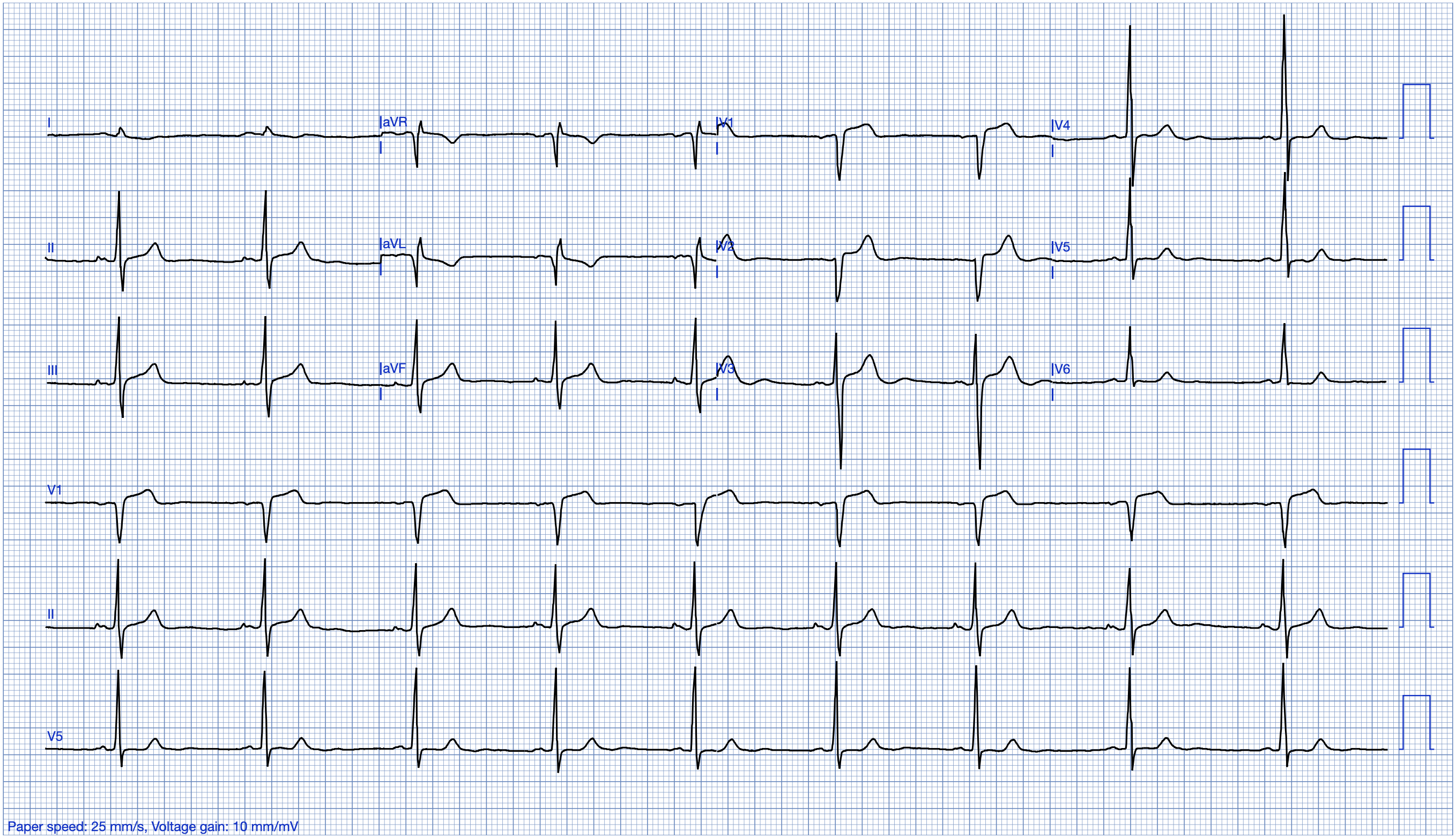
A man in his early 40s with no past medical history experienced acute onset crushing chest pain and dyspnea. The chest pain radiated into his left arm, and there was finger tip numbness. He rated it 10 out of 10. He took aspirin 325 mg and called EMS. The EMS report describes him as diaphoretic and clammy with extreme anxiety. His ECG is shown.
ECG 1
What do you think?
The Queen of Hearts calls this negative for OMI, but the raw output is 0.48. The model output ranges from 0 to 1 where 0 is no evidence for OMI and 1 is maximal confidence for OMI. The threshold for positivity is 0.5. So 0.48 is extremely close to being positive, and even a few subtle changes in digitization could conceivably swing this above 0.5. In a patient with a classic history for OMI (i.e. very high pre-test probability), this is not reassuring at all.
____
New PMcardio for Individuals App 3.0 now includes the latest Queen of Hearts model and AI explainability (blue heatmaps)! Download now for iOS or Android. (Dr. Smith is a shareholder in Powerful Medical.)
____
This is a difficult ECG, but knowing the history my interpretation was precordial swirl. Specifically, there is STE with hyperacute T waves in V1 with flat, very subtly downsloping STD in V6. The inferior T waves are also generous in size, with what appears to be reciprocal STD in the high lateral leads and ischemic down-up T waves especially in lead I.
Documentation indicates that the symptoms got "better" in the 15 minutes prior to arrival, but it does not say symptoms were RESOLVED. Ischemic chest pain is either resolved or unresolved. "Better" is almost meaningless. Initial high sensitivity troponin I (hsTnI) was 5 ng/L (ref. < 35 ng/L). The patient did not receive any nitro.
The next update an hour later says he had return of chest pain prompting repeat ECG, shown below.
ECG 2

Overall, pretty similar looking but probably a little improved. The ECG is irrelevant at this point, since the history is essentially diagnostic for OMI. And in a patient with stuttering symptoms, emergent angiography is the appropriate management. Cardiology recommended nitroglycerin, but none was apparently given. Repeat hsTnI rose from 5 ng/L to 64 ng/L.
Documentation indicates return of symptoms two hours later. It is not clear what happened during the prior symptomatic episode. Repeat ECG was very similar, and the patient received 0.4 mg sublingual nitroglycerin with "improvement" in pain. Third hsTnI resulted at 269 ng/L. At this point, he was diagnosed with "NSTEMI" and started on continuous heparin infusion.
A few hours later still, the patient had return of symptoms and received two additional doses of sublingual nitroglycerin with "slight improvement." HsTnI resulted at 1013 ng/L. This was in the middle of the night. The on-call interventional cardiologist recommended cardiac catheterization in the morning. This overt departure from guideline recommended management (immediate angiography for medically refractory chest pain) is reflective of usual care in the real world. About 1 out of every 15 patients with refractory angina is treated in accordance with guidelines.
The next day, the patient went for angiography. He was the second case the following day, after an elective outpatient procedure. (Why rush for an NSTEMI?) His door to angiography time was 13 hours and 54 minutes.
Shown below is a selected projection from his angiogram. The diagnostic finding is visualized here. See if you can find it. (I overlooked it until it was pointed out to me.)
Video: AP Cranial Angiogram
Here is a narrated video explaining the angiogram:
Video: AP Cranial Angiogram shown above, now with slow motion freeze frame.
WILLY — I added the video below. For YOU to decide if you want to use it! — :) Ken
I have personally never seen isolated septal perforator OMI before. But it makes perfect sense given the finding of precordial swirl on ECG 1. Swirl indicates a rightward vector of injury (since the septum is rightward relative to the bulk of LV mass). This was not felt to be a suitable target for PCI. I have never personally seen intervention to a septal perforator, but I was able to find a few case reports. This is extremely rarely done for a variety of reasons (technical difficulty, smaller caliber vessel, mechanical stress on stent from intramyocardial course, etc.) He was managed with dual antiplatelet therapy.
HsTnI peaked at 22,286 ng/L and echocardiogram showed LVEF 49% with septal akinesis.
Discussion
First, what a fascinating case! I asked several experienced cardiologists, and no one I talked to had ever seen an isolated septal perforator infarct. The remaining coronary vessels were essentially angiographically normal other than subjectively slow flow which may be suggestive of microvascular dysfunction. Thus, the etiology of the infarct is not entirely clear. Classic plaque rupture is probably the most statistically likely explanation, but it is a bit unusual his remaining vessels did not show any plaque. (However, in patients with positive remodeling, CAD may fail to produce luminal narrowing rendering it essentially invisible on invasive angiography.) Embolism is also possible, although there was no apparent embolic source.
Second, what a shame that he received such delayed angiography. In this case, the delay MAY not have changed his outcome since no intervention was performed. But it is also possible something useful could have been done if the angiogram had been done 13 hours earlier. Perhaps at that time a large thrombus could have been aspirated. There is no telling how things changed as he completed his infarct in the hospital.
More to the point, the fact that no intervention was performed does not mean delaying the angiogram was reasonable BEFORE that information was known. This could equally well have been a proximal LAD lesion.
But it is easy to see why cases like this reinforce the biases of the physicians. Another NSTEMI where delaying cath did not appear to matter to the eventual outcome. That is how a STEMI disciple thinks of it. And in the world of STEMI, this is considered normal care. There is no way for this to be flagged as a systems failure for process improvement. Even though he had a large infarct (with classic symptoms), lost his septum, and experienced reduction in LVEF.
No comments:
Post a Comment